Testing Capabilities
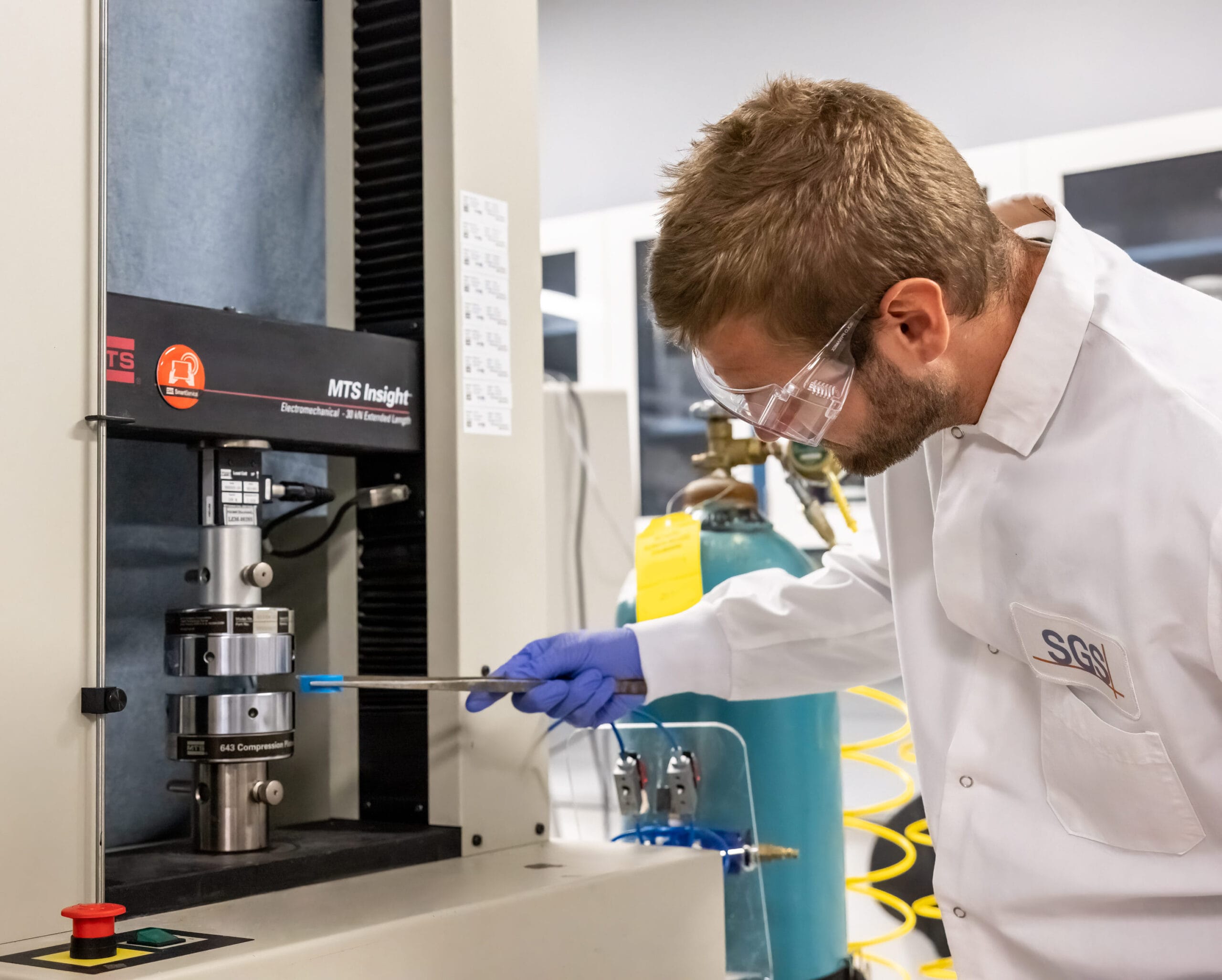
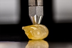
We support a wide range of materials and physical testing capabilities, as well as chemical analysis, leveraging innovative state-of-the art technologies.
We have a proven track record of solving complex problems for our clients.
We help you avoid unnecessary costs and unintended consequences for your critical projects and products so that you can go to market with peace of mind.
What are you looking for?