
Microscopy
The TEM is capable of examining single columns of atoms at magnifications that exceed optical microscopy (OM) by tens of thousands of times.
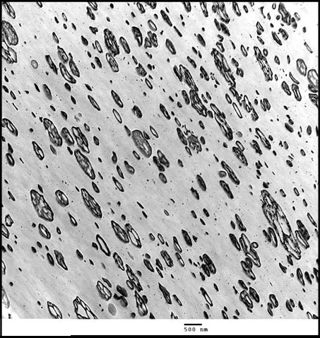
A concentrated beam of electrons passes through a very thin sample. Based on electron density the various areas of the sample interact with the beam differently. The resulting image represents the gradient areas of electron density.
Approaches
Transmission Electron Microscopy Analysis is a great approach for documenting the phase distribution of copolymers and blends.
Many samples have sufficient contrast in an as-prepared state. However, to obtain sufficient contrast with some specimens, a staining technique is utilized. For example, unsaturated components within a matrix are stained with ruthenium tetroxide. This method will reveal the rubber phase distribution in a polystyrene matrix.
Ultra thin specimens are prepared using a microtome or a cryo microtome on polymer pieces or embedded specimens.
Energy dispersion spectroscopy (EDS) can be used along with the TEM imaging to provide elemental compositions of sample features.
Sample Considerations
TEM specimens are typically very thin, approximately 0.1 microns in thickness. Specimens are acquired from regions of the sample based on the goal you hope to achieve through TEM testing.
Contact an expert to talk through your specific sample considerations
Experience
Our experience with Transmission Electron Microscopy Analysis includes:
- Phase morphology
- Dispersion of polymer blends
- Material selection
- Quality control
- Failure analysis
- Competitive product analysis
- Cellular structure of dispersed phases
- Nanoscale particle incorporation
- Metalized layer coherence and thickness analysis in laminated films