Parkinson’s disease, spinal cord injury, and chronic pain are devastating neurological conditions that are difficult for physicians to treat. The trouble lies in the inability of the physician to bypass or heal damaged neurons. A device capable of stimulating neural tissue and recording the subsequent activity simultaneously could potentially help repair damaged neural tissue in patients with paralysis due to spinal cord injury. A device of this nature would have to be highly flexible and biocompatible.
New Approach to Implants
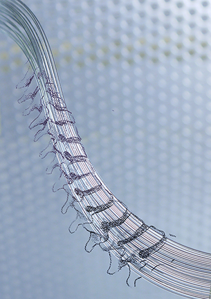
Graduate student Chi (Alice) Lu, of the Polymer Science and Technology Program at MIT is the designer of highly flexible neural probes. The probes are made using a polycarbonate (PC) and cyclic olefin copolymer (COC) core. The PC fiber is approximately 1.5 inches thick and is pulled using thermal drawing, until it reaches a thickness of 400-1,100 microns (µm). The fiber is then etched to reach a final thickness of 180-230 µm. The flexibility of the electrodes is extremely high, and they can be tied into a knot. Tests showed the probes performed well under the stressful environment and natural movements of the body. The design also includes a waveguide for stimulating optically and conductive polyethylene (CPE) electrodes to record data.
The experimental procedure included testing the neural implant in mice whose neurons responded to blue light. The mice expressed the protein channelrhodopsin 2 (ChR2), which is light-sensitive and caused the neural response. A neuronal response is generated by shining a light on the spinal cord; the response is recorded. Lu said:
The next step will be a chronic study. I think the challenge will be how to implant even a very flexible device without paralyzing the animal. We are just interfacing something harder than the tissue with the body. And then when the animal moves, the device has to follow this movement organically otherwise it will potentially paralyze the animal. So this will be another challenge, how to find a good spot for stimulation and recording, but not damage the tissue.
Existing Implantable Devices
Currently, deep brain and spinal cord stimulation uses antiquated electrode technology. The implants are not biocompatible, and their inflexibility causes mechanical damage to the surrounding tissue. The materials used for the implants elicit an adverse response from the neurological tissue, which often leads to the encapsulation of the implant in scar tissue, which can render the implant useless.
Another potential problem is the deposition of the electrode material into the neural tissue, which is caused when an electrical current passes through the probe, etching off material. However, due to advances in materials science and fabrication techniques, new and better neurological implants are making the news.
Probe Potential
There are limitations as far as medical applications are concerned. Human neurons are not light-sensitive, with the exception of optical neurons. Polina Anikeeva, AMAX assistant professor in materials science and engineering at MIT, would like to see this work taken a step further, aiding in the development of flexible biomimetic optoelectronic neuroprosthetics. This would allow the device to communicate with nerves for use with limb prosthetic devices.
Lu’s neural device may not be ready for human trials, but it is a step in the right direction. Biocompatibility is critical for an implantable neural device to function properly, if at all. Flexibility is also vital in an area as sensitive and dynamic as the central nervous system. Chronic studies will give us a better idea of how well these novel neural devices will work in the brain and spinal cord.
Image by Chi (Alice) Lu and Polina Anikeeva.
Source: “Flexible Polymer Probes and Magnetic Nanoparticles Promise Breakthroughs for Treating Paralysis, Brain Disease,” by Denis Paiste, www.phys.org, September 3, 2014.
Source: “Magnetic Nanoparticles and Polymer Fiber Probes for Treating Neurological Disorders,” by Alessandro Pirolini, www.azom.com, September 4, 2014.
Source: “Stimulating Nerves With Light,” by Denis Paiste, mpc-www.mit.edu, August 31, 2014.
Source: “Flexible Probes for Spinal Cord Integration,” Materials Views, www.materialsviews.com, September 5, 2014.
Source: “Biocompatible Materials for Optoelectronic Neural Probes: Challenges and Opportunities,” by Polina Ankeeva, National Academy of Engineering, www.nae.edu, Winter 2013.
Source: “Polymer Fiber Probes Enable Optical Control of Spinal Cord and Muscle Function In Vivo,” by Chi Lu, et al., Advanced Functional Materials, August 26, 2014, DOI: 10.1002/adfm.201401266.
